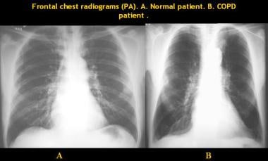
Bilateral Emphysematous Lung Fields

Chronic Obstructive Lung Disease Copd Emphysema

Chest X Ray Showing Predominant Emphysematous Changes Along With A Download Scientific Diagram
Emphysema Imaging Practice Essentials Radiography Computed Tomography
Teaching File Radiologic Anatomy Of The Lung

Bilateral Pulmonary Hyperaeration Metabolic Disorders

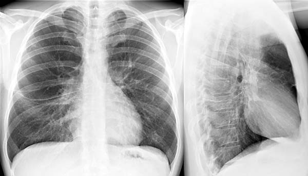
Lung Hyperinflation Radiology Reference Article Radiopaedia Org

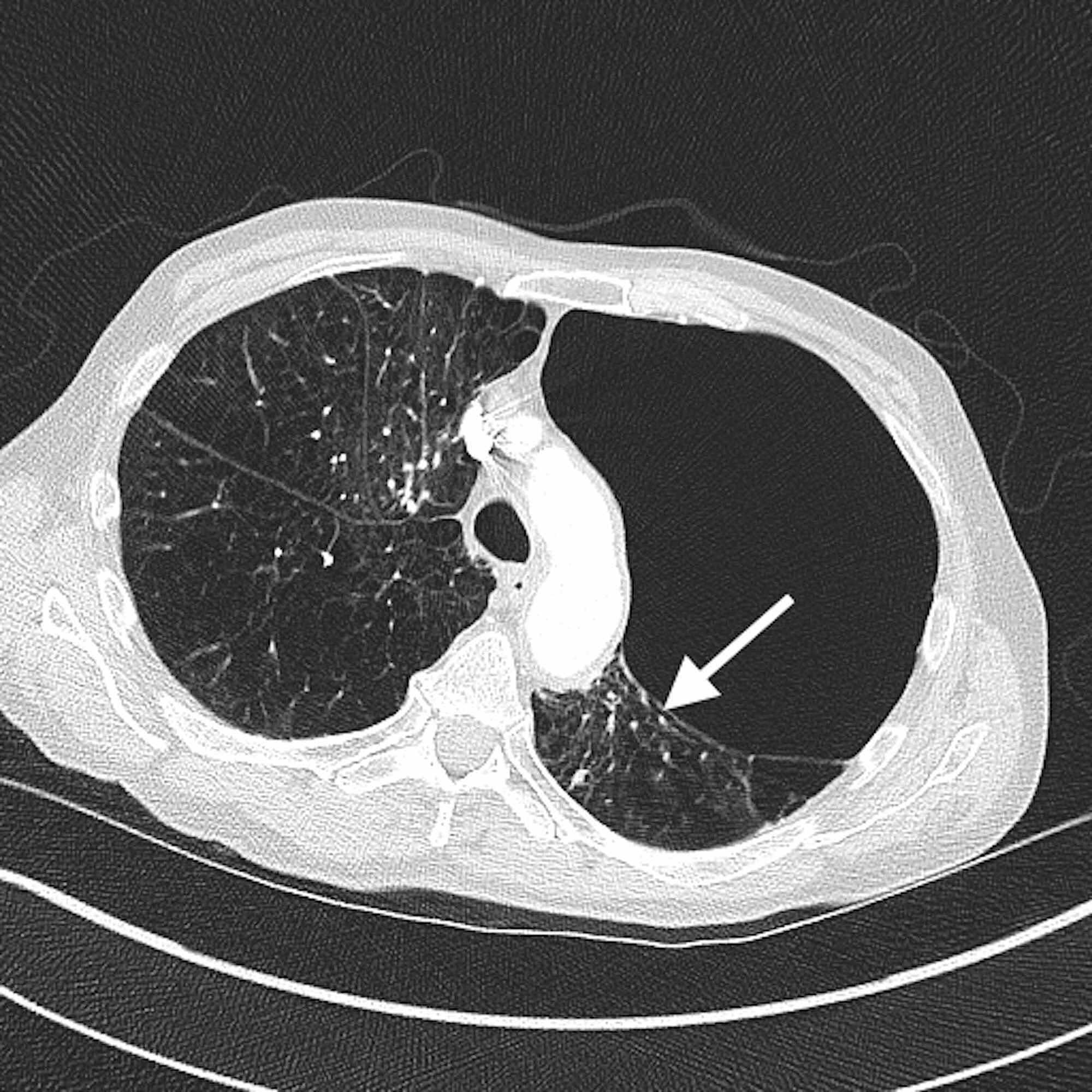
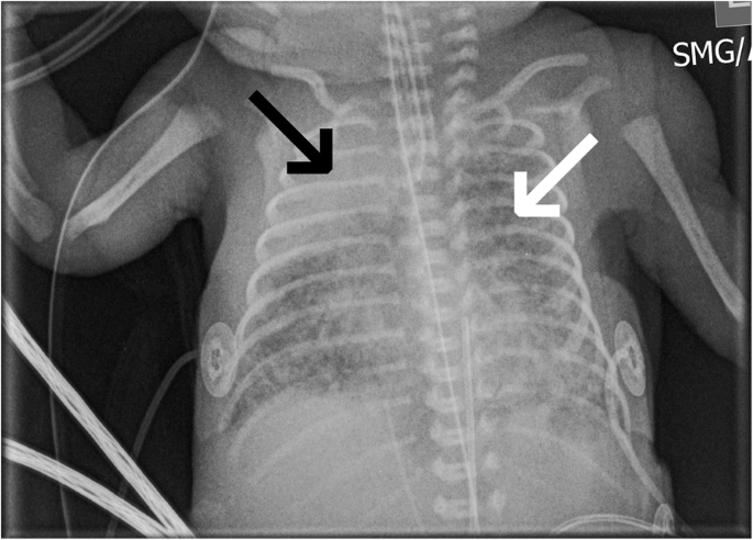
Paraseptal emphysema refers to inflammation and tissue damage to the distal airways and alveolar sacs near the outer boundaries of the lungs.
Bilateral emphysematous lung fields. Therefore treatment is centered on symptom management. It measures how much air you can blow out of your lungs in 1 second. A collapsed lung is an uncommon but serious condition that can be life threatening for people in advanced stages of emphysema. Prior to listening over any one area of the chest remind yourself which lobe of the lung is heard best in that region.
Mild emphysema is the early development of emphysemic symptoms associated with the presence of chronic obstructive pulmonary disease the condition is induced by the deterioration of the air sacs within one s lungs. Hyperinflated lungs can be caused by blockages in the air passages or by air sacs that are less elastic which interferes with the expulsion of air from the lungs. While more common types of emphysema impair major airway structures and disrupt normal airflow paraseptal emphysema is unlikely to cause noticeable breathing problems in its initial stages. Where in the lungs does emphysema show up.
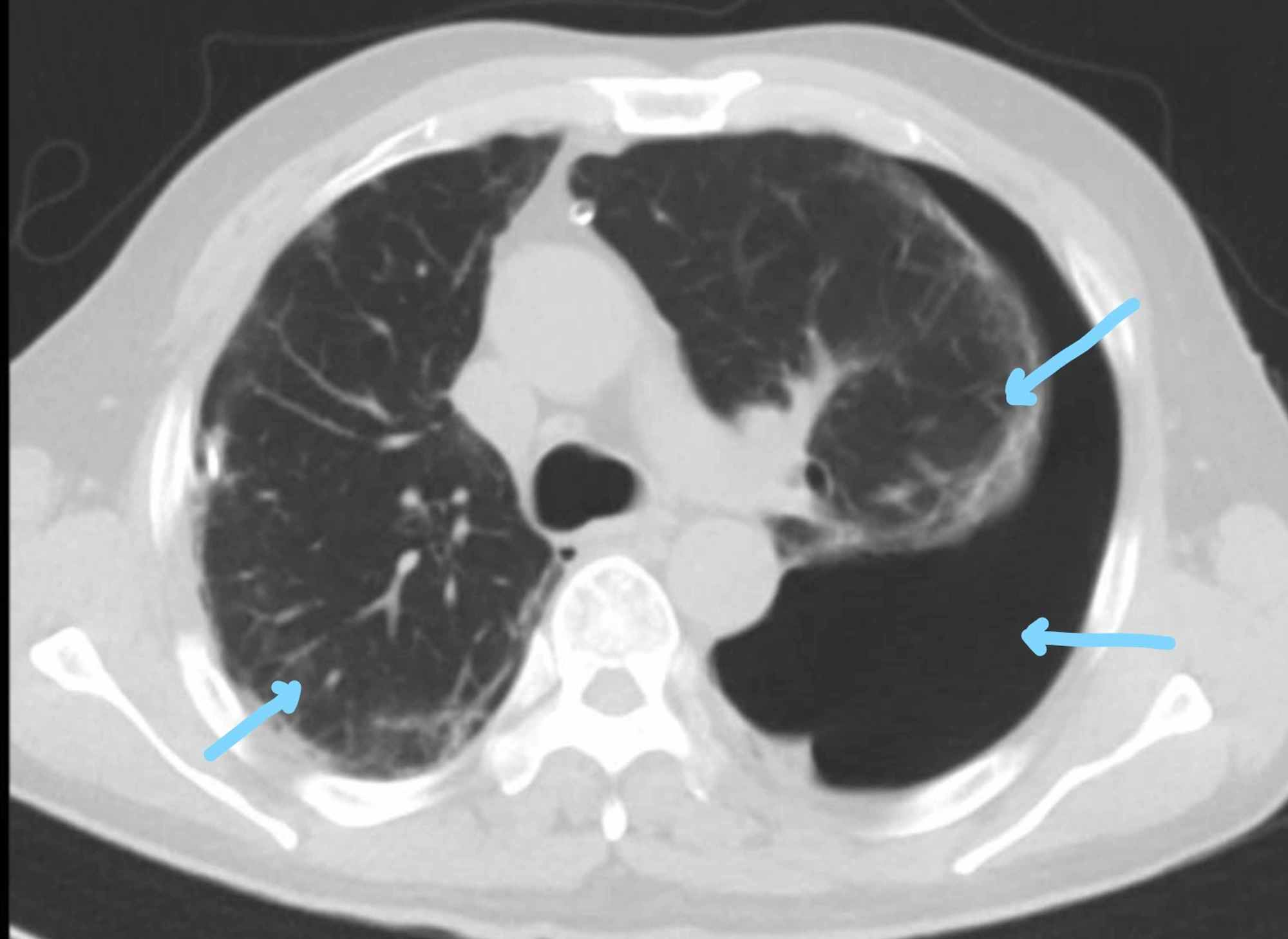
Apical predominant bullae which are most commonly associated with emphysema in the surrounding lung parenchyma. While we encourage individuals to share their personal experiences. Right middle lobe heard in right axilla. Hyperinflated lungs are often seen in people with chronic obstructive pulmonary disease copd a disorder that includes emphysema.
Emphysema is a lung condition that causes shortness of breath. Over time the inner walls of the air sacs weaken and rupture creating larger air spaces instead of many small ones. This reduces the surface area of the lungs and in turn the amount of oxygen that. Lingula in left axilla.
Although this condition commonly occurs in the tissue of the neck or chest wall it can develop in. Subcutaneous emphysema is a type of lung disease where air or gas gets under your skin tissue. Is there any way to determine cause of reduction in lung function. Doctors call this.
In people with emphysema the air sacs in the lungs alveoli are damaged. This is a set of guidelines established by the global initiative for chronic obstructive lung disease gold. Basilar predominant disease may be associated with alpha 1 antitrypsin deficiency or iv drug use. Lower lobes occupy the bottom 3 4 of the posterior fields.
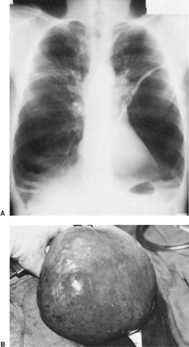
Holes in the lungs known as bullae can. The presence of mild symptoms is indicative of a progressive condition that has no cure. It is not our intention to serve as a substitute for medical advice and any content posted should not be used for medical advice diagnosis or treatment.

Chronic Obstructive Diseases Of The Lung Part 3
Bullous And Bleb Diseases Of The Lung Thoracic Key

Persistent Pulmonary Interstitial Emphysema In A Case Of Langerhans Cell Histiocytosis

Silicosis In The Form Of Progressive Massive Fibrosis A Diagnostic Challenge

Cardiothoracic Imaging Bilateral Emphysema Chronic Obstructive Pulmonary Disease Enlarged Heart Pulmonary

Imaging From A 64 Year Old Man With Cpfe A Hrct Of Bilateral Upper Download Scientific Diagram

Cannabis Bong Smoking Induced Pneumomediastinum And Subcutaneous Emphysema Radiology Case Radiopaedia Org

Pulmonary Interstitial Emphysema Radiology Case Radiopaedia Org
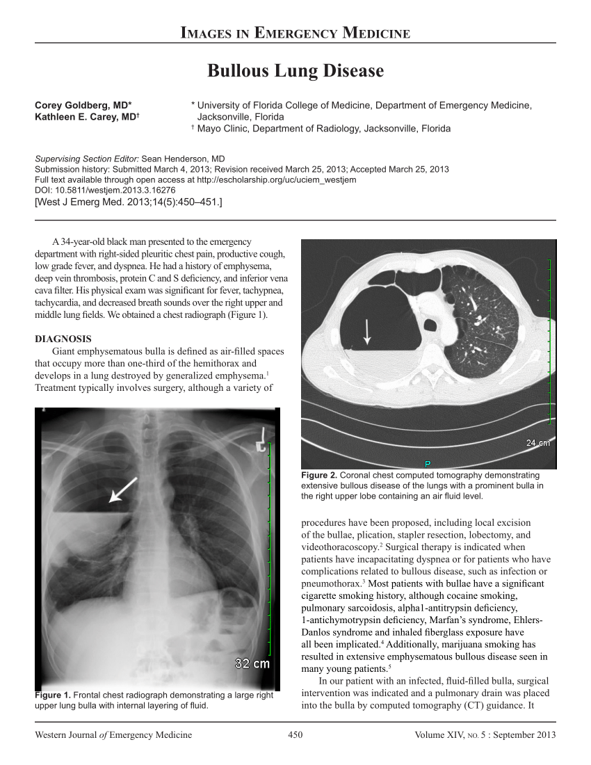
Pdf Bullous Lung Disease

Pin By Kathy I On Radiology Copd Lunges Radiology

Emphysema With Images Copd Lung Disease Lunges

E X20rfsg84elm

Hyperlucent Lung Syndrome Associated With Persistent Foramen Ovale And Bilateral Bullous Emphysema Consultant360
Perioperative Management Of Esophagectomy In A Patient Who Previously Underwent Bilateral Lung Transplantation Springerlink
Pdf Emphysema And Copd In A Young Woman

Regional Ventilation And Perfusion After Lung Transplantation In Patients With Emphysema Nejm
Article Fulle Text

Jaypeedigital Ebook Reader
Https Encrypted Tbn0 Gstatic Com Images Q Tbn 3aand9gcs Frib0buwx1mhxnpsgaz8qstkmod16edyuebeh3g Usqp Cau
Pulmonary Emphysema Radiology Reference Article Radiopaedia Org

Ct Scans Showing Tracheomegaly A Bilateral Upper Lobe Emphysema B Download Scientific Diagram

Subcutaneous Emphysema Radiology Case Radiopaedia Org Radiology Radiology Imaging Subcutaneous Emphysema
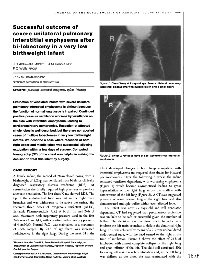
Pdf Successful Outcome Of Severe Unilateral Pulmonary Interstitial Emphysema After Bi Lobectomy In A Very Low Birthweight Infant

Pdf Percutaneous Evacuation Of Diffuse Pulmonary Interstitial Emphysema By Lung Puncture In A Baby With Extremely Low Birth Weight A Case Report

Hyperlucent Thorax Radiology Key
Cureus Vanishing Lung Syndrome An Idiopathic Bullous Emphysema Mimicking Pneumothorax
Cureus Bilateral Hemopneumothorax In Covid 19

Pediatric Radiology Pediatric Radiology Radiology Chronic Obstructive Pulmonary

Pdf Imaging Diagnosis Of Interstitial Pneumonia With Emphysema Combined Pulmonary Fibrosis And Emphysema
Pulmonary Nm V Q Nucmedresource Com
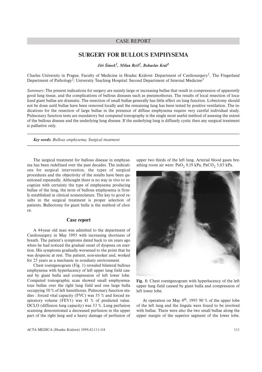
Pdf Surgery For Bullous Emphysema

File Barrowchest Jpg Wikipedia The Free Encyclopedia Radiology Imaging Radiology X Ray
Https Www Sciencedirect Com Science Article Pii S0025712516322684 Pdf Md5 672111efc3c683ba3e31b19a0766d59d Pid 1 S2 0 S0025712516322684 Main Pdf

Emergency Department Diagnosis Of Idiopathic Pneumoparotitis With Cervicofacial Subcutaneous Emphysema In A Pediatric Patient The Western Journal Of Emergency Medicine
Pictorial Review Of Non Traumatic Thoracic Emergencies In The Pediatric Population Springerlink
Atelectasis Wikipedia

Let S So Systematically On This One 1 Trachea Deviated To Right 2 Calcified Group Of Cervical Lymph Node 3 Pleural Thicking Trachea Lymph Nodes Cervical

Flow Chart Of Patients Included In The Study Bilateral Emphysema In Download Scientific Diagram

Hrct Showing Emphysema Changes In The Upper Lobes A Emphysema And Download Scientific Diagram

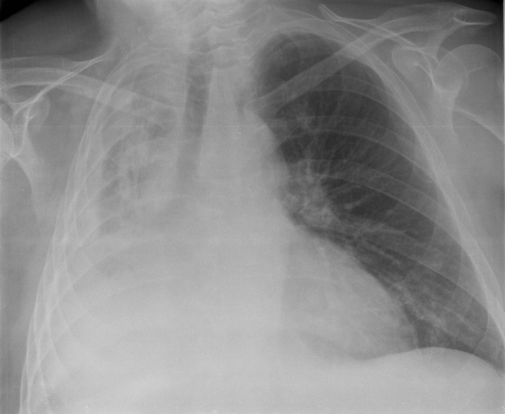
Omplete White Out Of A Hemithorax On The Chest X Ray Has A Limited Number Of Causes The Differential Diagnosis Can B Renal Cell Radiology Renal Cell Carcinoma

Chest Including Lungs And Mediastinum Radiology Key

Chronic Obstructive Pulmonary Disease Copd Represents A Spectrum Of Obstructive Airway Diseases It Includes Two Key Radiology Radiology Imaging Pulmonology

